Radiographic features and molecular subtypes association of breast cancer in women younger than 40 years old
DOI:
https://doi.org/10.46475/asean-jr.v25i1.174Keywords:
Breast cancer, Breast imaging, Histopathological type, molecular subtype, Young womenAbstract
Background: Breast cancer is the most commonly diagnosed cancer and the leading cause of cancer death among females, particularly in women under the age of 40 years. However, early detection of breast cancer in this population remains challenging and it tends to present at a later stage with poorer prognosis.
Objective: To review mammographic and ultrasonographic findings, pathological features and molecular subtypes of breast cancer in younger than 40-year-old patients diagnosed in King Chulalongkorn Memorial Hospital and to determine which radiological characteristics are associated with molecular subtypes.
Materials and Methods: The study included 278 patients aged under 40 years who were diagnosed with breast cancer and underwent mammographic and ultrasonographic studies between January 2009 and December 2019. A retrospective review of mammographic and ultrasonographic findings, histopathological reports as well as biological markers were made. The association of radiological characteristics and molecular subtypes was analyzed by SPSS.
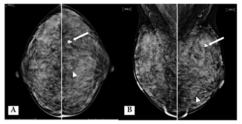
Results: In the 278 patients, the most common clinical presentation was palpable mass (268, 96.4%). The common mammographic findings were irregular shape mass (196, 77.8%) with hyperdensity (114, 45.2%) and an obscured margin (99, 39.3%). Presenting of microcalcification is not frequent (122, 48.4%). We found 27 patients with normal mammograms which were later detected in ultrasounds as 25 masses, 1 intraductal lesion and 1 focal duct change. The predominant ultrasonographic features were irregular shape mass (257, 91.5%), an angular margin (89, 31.7%), hypoechogenicity (198, 70.5%), no posterior feature (210, 74.7%) and internal vascularity (170, 60.5%). These radiological characteristics were classified as BI-RADS 5 in 194 lesions (69%). The most common histopathological type was mixed-type carcinoma (143, 50.9%), followed by invasive ductal carcinoma (114, 40.6%). Luminal B was the mostly found in this study (86, 30.6%). The patients frequently presented with stage IIA (91, 32.7%) while 15 patients were detected with an advanced stage at the first presentation. We found that triple negative, HER 2 overexpression and luminal B subtypes were associated with an obscured mass on mammography (p 0.048). Luminal B and HER 2 overexpression subtypes were also associated with the presence of fine pleomorphic microcalcification (p <0.001).
Conclusion: In this study, we found an association of the mass margin and suspicious calcification morphology on mammography with molecular subtypes. It would be helpful for further clinical management in young patients. The knowledge can be used for planning appropriate treatments according to molecular subtypes which are associated with these characteristics. However, the precision of cancer treatment is still based on the tissue diagnosis.
Downloads
Metrics
References
Bray F, Ferlay J, Soerjomataram I, Siegel RL, Torre LA, Jemal A. Global cancer statistics 2018: GLOBOCAN estimates of incidence and mortality worldwide for 36 cancers in 185 countries. CA Cancer J Clin 2018;68:394-424. doi: 10.3322/caac.21492. DOI: https://doi.org/10.3322/caac.21492
Paluch-Shimon S, Pagani O, Partridge AH, Bar-Meir E, Fallowfield L, Fenlon D, et al. Second international consensus guidelines for breast cancer in young women (BCY2). Breast 2016;26:87-99. doi: 10.1016/j.breast.2015.12.010. DOI: https://doi.org/10.1016/j.breast.2015.12.010
Assi HA, Khoury KE, Dbouk H, Khalil LE, Mouhieddine TH, El Saghir NS. Epidemiology and prognosis of breast cancer in young women. J Thorac Dis 2013;5 Suppl 1:S2-8. doi: 10.3978/j.issn.2072-1439.2013.05.24.
Anders CK, Johnson R, Litton J, Phillips M, Bleyer A. Breast cancer before age 40 years. Semin Oncol 2009;36:237-49. doi: 10.1053/j.seminoncol.2009.03.001. DOI: https://doi.org/10.1053/j.seminoncol.2009.03.001
Durhan G, Azizova A, Önder Ö, Kösemehmetoğlu K, Karakaya J, Akpınar MG, et al. Imaging Findings and Clinicopathological Correlation of Breast Cancer in Women under 40 Years Old. Eur J Breast Health 2019;15:147-52. doi: 10.5152/ejbh.2019.4606. DOI: https://doi.org/10.5152/ejbh.2019.4606
Gillman J, Batel J, Chun J, Schwartz S, Moy L, Schnabel F. Imaging and clinicopathologic characteristics in a contemporary cohort of younger women with newly diagnosed breast cancer. Cancer Treat Res Commun 2016;9:35-40. DOI: https://doi.org/10.1016/j.ctarc.2016.06.006
American Cancer Society. Cancer facts and figures 2019. Atlanta: American Cancer Society; 2019.
Hortobagyi GN, Connolly JL, D’Orsi CJ, Edge SB, Mittendorf EA, Hope S, et al. Breast. In: Amin MB, Edge S, Greene F, Byrd DR, Brookland RK, Washington MK, et al, editors. 8th ed. New York: Springer; 2017. p. 589-628.
Lakhani SR, Ellis IO, Schnitt SJ, Tan PH, van de Vijver MJ, editors. WHO classification of tumours of the breast: WHO Classification of Tumours, 4th ed. Volume 4. Lyon: IARC Press; 2012.
D’Orsi CJ, Sickles EA, Mendelson EB, Morris EA. ACR BI-RADS® Atlas, Breast Imaging Reporting and Data System. Reston(VA): American College of Radiology; 2013.
Davey MG, Brennan M, Ryan ÉJ, Corbett M, Abd Elwahab S, Walsh S, et al. Defining clinicopathological and radiological features of breast cancer in women under the age of 35: an epidemiological study. Ir J Med Sci 2020;189:1195-202. doi: 10.1007/s11845-020-02229-z. DOI: https://doi.org/10.1007/s11845-020-02229-z
Shoemaker ML, White MC, Wu M, Weir HK, Romieu I. Differences in breast cancer incidence among young women aged 20-49 years by stage and tumor characteristics, age, race, and ethnicity, 2004-2013. Breast Cancer Res Treat 2018;169:595-606. doi: 10.1007/s10549-018-4699-9. DOI: https://doi.org/10.1007/s10549-018-4699-9
Kim, J, Jang M, Kim SM, Yun BL, Lee JY, Kim EK, et al. Clinicopathological and imaging features of breast cancer in Korean women under 40 years of age. J Korean Soc Radiol 2017;76:375-85. DOI: https://doi.org/10.3348/jksr.2017.76.6.375
Eugênio DSG, Souza JA, Chojniak R, Bitencourt AGV, Graziano L, Marques EF. Breast cancer diagnosed before the 40 years: imaging findings and correlation with histology and molecular subtype. Appl Cancer Res [Internet]. 2017 [cited 2024 Feb 19];37(16). Available from: https://appliedcr.biomedcentral.com/articles/10.1186/s41241-017-0019-7#article-info DOI: https://doi.org/10.1186/s41241-017-0019-7
Collins LC, Marotti JD, Gelber S, Cole K, Ruddy K, Kereakoglow S, et al. Pathologic features and molecular phenotype by patient age in a large cohort of young women with breast cancer. Breast Cancer Res Treat 2012;131:1061-6. doi: 10.1007/s10549-011-1872-9. DOI: https://doi.org/10.1007/s10549-011-1872-9
Tirada N, Aujero M, Khorjekar G, Richards S, Chopra J, Dromi S, et al. Breast cancer tissue markers, genomic profiling, and other prognostic factors: A primer for radiologists. RadioGraphics 2018;38:1902-20. doi: 10.1148/rg.2018180047. DOI: https://doi.org/10.1148/rg.2018180047
Bullier B, MacGrogan G, Bonnefoi H, Hurtevent-Labrot G, Lhomme E, Brouste V, et al. Imaging features of sporadic breast cancer in women under 40 years old: 97 cases. Eur Radiol 2013;23:3237-45. doi: 10.1007/s00330-013-2966-z. DOI: https://doi.org/10.1007/s00330-013-2966-z
Erić I, Petek Erić A, Kristek J, Koprivčić I, Babić M. Breast cancer in young women: Pathologic and immunohistochemical features. Acta Clin Croat 2018;57:497-502. doi: 10.20471/acc.2018.57.03.13. DOI: https://doi.org/10.20471/acc.2018.57.03.13
Expert Panel on Breast Imaging; Mainiero MB, Moy L, Baron P, Didwania AD, diFlorio RM, , et al. ACR Appropriateness Criteria® Breast Cancer Screening. J Am Coll Radiol 2017;14(11 Suppl):S383-90. doi: 10.1016/j.jacr.2017.08.044. DOI: https://doi.org/10.1016/j.jacr.2017.08.044

Downloads
Published
How to Cite
Issue
Section
License
Copyright (c) 2024 The ASEAN Journal of Radiology

This work is licensed under a Creative Commons Attribution-NonCommercial-NoDerivatives 4.0 International License.
Disclosure Forms and Copyright Agreements
All authors listed on the manuscript must complete both the electronic copyright agreement. (in the case of acceptance)


