Comparison between the use of one and two CT scans for attenuation correction of rest-stress myocardial perfusion SPECT with Tc-99m sestamibi
DOI:
https://doi.org/10.46475/asean-jr.v25i2.895Keywords:
CT-based attenuation correction, Gated SPECT, Left ventricle function, Myocardial perfusion SPECT, Tc-99m sestamibiAbstract
Background: The standard protocol is to use separate computed tomography (CT) scans acquired during rest and stress for attenuation correction (AC) of myocardial perfusion (MP) single photon emission computed tomography (SPECT) imaging. Recently, there have been attempts to reduce the radiation dose by using one CT instead of two CTs.
Objective: To compare between the use of one and two CTs for AC of rest-stress MP SPECT with Tc-99m sestamibi in quantification of MP and left ventricle (LV) function.
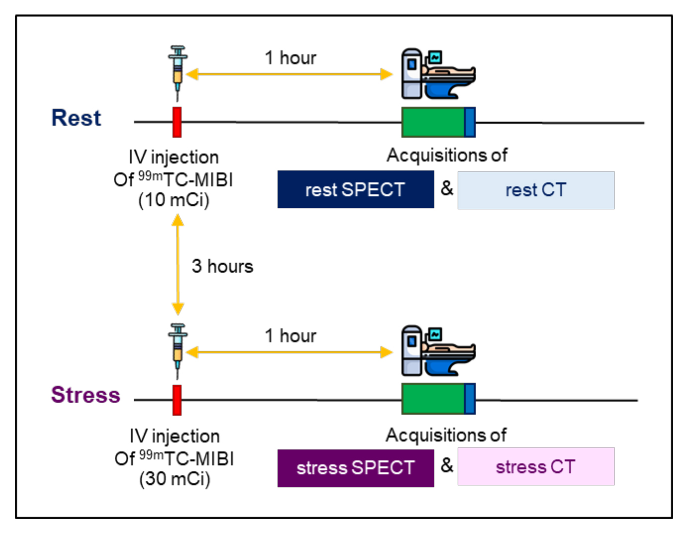
Materials and Methods: Gated rest-stress MP SPECT images of 107 patients were reprocessed using 3 different AC methods: 1) rest CT for AC of rest SPECT and stress CT for AC of stress SPECT (2CT); 2) rest CT for AC of both rest and stress SPECT (1CT-rest); and 3) stress CT for AC of both rest and stress SPECT (1CT-stress). SPECT images obtained from 2CT and 1CT were used for quantification of MP values and LV function values. The values from 2CT and 1CT were compared.
Results: The MP values of 2CT and 1CT showed a strong correlation (r≥0.712) and they did not differ significantly (p=0.106 to 0.931). In contrast, the LV function values of 2CT and 1CT exhibited a very strong correlation (r≥0.960), but they differ significantly (p=<0.001 to 0.004).
Conclusions: The use of one and two CTs for AC in rest-stress MP SPECT with Tc-99m sestamibi can be interchanged for the quantification of MP, but not for the quantification of LV function.
Downloads
Metrics
References
Bateman TM, Cullom SJ. Attenuation correction single-photon emission computed tomography myocardial perfusion imaging. Semin Nucl Med 2005;35:37-51. doi: 10.1053/j.semnuclmed.2004.09.003. DOI: https://doi.org/10.1053/j.semnuclmed.2004.09.003
Jaarsma C, Leiner T, Bekkers SC, Crijns HJ, Wildberger JE, Nagel E, et al. Diagnostic performance of noninvasive myocardial perfusion imaging using single-photon emission computed tomography, cardiac magnetic resonance, and positron emission tomography imaging for the detection of obstructive coronary artery disease: a meta-analysis. J Am Coll Cardiol 2012;59:1719-28. doi: 10.1016/j.jacc.2011.12.040. DOI: https://doi.org/10.1016/j.jacc.2011.12.040
Rischpler C, Nekolla S, Schwaiger M. PET and SPECT in heart failure. Curr Cardiol Rep 2013;15:337. doi: 10.1007/s11886-012-0337-z. DOI: https://doi.org/10.1007/s11886-012-0337-z
Cremer P, Hachamovitch R, Tamarappoo B. Clinical decision making with myocardial perfusion imaging in patients with known or suspected coronary artery disease. Semin Nucl Med 2014;44:320-9. doi: 10.1053/j.semnuclmed.2014.04.006. DOI: https://doi.org/10.1053/j.semnuclmed.2014.04.006
Kostkiewicz M. Myocardial perfusion imaging in coronary artery disease. Cor et Vasa 2015;57:e446-52. DOI: https://doi.org/10.1016/j.crvasa.2015.09.010
Lehner S, Nowak I, Zacherl M, Brosch-Lenz J, Fischer M, Ilhan H, et al. Quantitative myocardial perfusion SPECT/CT for the assessment of myocardial tracer uptake in patients with three-vessel coronary artery disease: initial experiences and results. J Nucl Cardiol 2022;29:2511-20. doi: 10.1007/s12350-021-02735-2. DOI: https://doi.org/10.1007/s12350-021-02735-2
Strauss HW, Miller DD, Wittry MD, Cerqueira MD, Garcia EV, Iskandrian AS, et al. Procedure guideline for myocardial perfusion imaging 3.3. J Nucl Med Tech 2008;36:155-61. doi: 10.2967/jnmt.108.056465. DOI: https://doi.org/10.2967/jnmt.108.056465
Czaja M, Wygoda Z, Duszańska A, Szczerba D, Głowacki J, Gąsior M, et al. Interpreting myocardial perfusion scintigraphy using single-photon emission computed tomography. Part 1. Kardiochir Torakochirurgia Pol 2017;14:192-9. doi: 10.5114/kitp.2017.70534. DOI: https://doi.org/10.5114/kitp.2017.70534
Abidov A, Germano G, Hachamovitch R, Slomka P, Berman DS. Gated SPECT in assessment of regional and global left ventricular function: an update. J Nucl Cardiol 2013;20:1118-43. doi: 10.1007/s12350-013-9792-1. DOI: https://doi.org/10.1007/s12350-013-9792-1
Go V, Bhatt MR, Hendel RC. The diagnostic and prognostic value of ECG-gated SPECT myocardial perfusion imaging. J Nucl Med 2004;45:912-21.
Slart RH, Tio RA, Zeebregts CJ, Willemsen A, Dierckx RA, De Sutter J. Attenuation corrected gated SPECT for the assessment of left ventricular ejection fraction and volumes. Ann Nucl Med 2008;22:171-6. doi: 10.1007/s12149-007-0100-5. DOI: https://doi.org/10.1007/s12149-007-0100-5
Bavelaar-Croon CD, Kayser HW, van der Wall EE, de Roos A, Dibbets-Schneider P, Pauwels EK, et al. Left ventricular function: correlation of quantitative gated SPECT and MR imaging over a wide range of values. Radiology 2000; 217:572-5. doi: 10.1148/radiology.217.2.r00nv15572. DOI: https://doi.org/10.1148/radiology.217.2.r00nv15572
Czaja MZ, Wygoda Z, Duszańska A, Szczerba D, Głowacki J, Gąsior M, et al. Myocardial perfusion scintigraphy-interpretation of gated imaging. Part 2. Kardiochir Torakochirurgia Pol 2018;15:49-56. doi: 10.5114/kitp.2018.74676. DOI: https://doi.org/10.5114/kitp.2018.74676
Hendel RC, Corbett JR, Cullom SJ, DePuey EG, Garcia EV, Bateman TM. The value and practice of attenuation correction for myocardial perfusion SPECT imaging: a joint position statement from the American Society of Nuclear Cardiology and the Society of Nuclear Medicine. J Nucl Cardiol 2002;9:135-43. doi: 10.1067/mnc.2002. DOI: https://doi.org/10.1067/mnc.2002.120680
Burrell S, MacDonald A. Artifacts and pitfalls in myocardial perfusion imaging. J Nucl Med Technol 2006;34:193-211 ; quiz 212-4.
Singh B, Bateman TM, Case JA, Heller G. Attenuation artifact, attenuation correction, and the future of myocardial perfusion SPECT. J Nucl Cardiol 2007;14:153-64. doi: 10.1016/j.nuclcard.2007.01.037. DOI: https://doi.org/10.1016/j.nuclcard.2007.01.037
Slart RH, Que TH, van Veldhuisen DJ, Poot L, Blanksma PK, Piers DA, et al. Effect of attenuation correction on the interpretation of 99mTc-sestamibi myocardial perfusion scintigraphy: the impact of 1 year's experience. Eur J Nucl Med Mol Imaging 2003;30:1505-9. doi: 10.1007/s00259-003-1265-3. DOI: https://doi.org/10.1007/s00259-003-1265-3
Masood Y, Liu YH, Depuey G, Taillefer R, Araujo LI, Allen S, et al. Clinical validation of SPECT attenuation correction using x-ray computed tomography-derived attenuation maps: multicenter clinical trial with angiographic correlation. J Nucl Cardiol 2005;12:676-86. doi: 10.1016/j.nuclcard.2005.08.006. DOI: https://doi.org/10.1016/j.nuclcard.2005.08.006
Garcia EV. SPECT attenuation correction: an essential tool to realize nuclear cardiology’s manifest destiny. J Nucl Cardiol 2007;14:16-24. doi: 10.1016/j.nuclcard.2006.12.144. DOI: https://doi.org/10.1016/j.nuclcard.2006.12.144
Huang JY, Huang CK, Yen RF, Wu HY, Tu YK, Cheng MF, et al. Diagnostic performance of attenuation-corrected myocardial perfusion imaging for coronary artery disease: a systematic review and meta-analysis. J Nucl Med 2016;57:1893-8. doi: 10.2967/jnumed.115.171462. DOI: https://doi.org/10.2967/jnumed.115.171462
Cerqueira MD, Allman KC, Ficaro EP, Hansen CL, Nichols KJ, Thompson RC, et al. Recommendations for reducing radiation exposure in myocardial perfusion imaging. J Nucl Cardiol 2010;17:709-18. doi: 10.1007/s12350-010-9244-0. DOI: https://doi.org/10.1007/s12350-010-9244-0
Einstein AJ. Multiple opportunities to reduce radiation dose from myocardial perfusion imaging. Eur J Nucl Med Mol Imaging 2013;40:649-51. doi: 10.1007/s00259-013-2355-5. DOI: https://doi.org/10.1007/s00259-013-2355-5
Ahlman MA, Suranyi P, Spicer KM, Gordon L. Comparison of one vs two CTs for attenuation correction of both rest and stress SPECT data for myocardial perfusion imaging with Tc-99m Tetrofosmin. J Nucl Med 2011;52:1129.
Ahlman MA, Nietert PJ, Wahlquist AE, Serguson JM, Berry MW, Suranyi P, et al. A single CT for attenuation correction of both rest and stress SPECT myocardial perfusion imaging: a retrospective feasibility study. Int J Clin Exp Med 2014;7:148-55.
Wells RG, Trottier M, Premaratne M, Vanderwerf K, Ruddy TD. Single CT for attenuation correction of rest/stress cardiac SPECT perfusion imaging. J Nuclear Cardiol 2018;25:616-24. doi: 10.1007/s12350-016-0720-z. DOI: https://doi.org/10.1007/s12350-016-0720-z
Fukami M, Tamura K, Nakamura Y, Nakatsukasa S, Sasaki M. Evaluating the effectiveness of a single CT method for attenuation correction in stress-rest myocardial perfusion imaging with thallium-201 chloride SPECT. Radiol Phys Technol 2020;13:20-6. doi: 10.1007/s12194-019-00540-8. DOI: https://doi.org/10.1007/s12194-019-00540-8
Slomka PJ, Berman DS, Germano G. Normal limits for transient ischemic dilation with 99mTc myocardial perfusion SPECT protocols. J Nucl Cardiol 2017;24:1709-11. doi: 10.1007/s12350-016-0582-4. DOI: https://doi.org/10.1007/s12350-016-0582-4
Preuss R, Weise R, Lindner P, Fricke E, Fricke H, Burchert W. Optimisation of protocol for low dose CT-derived attenuation correction in myocardial perfusion SPECT imaging. Eur J Nucl Med Mol Imaging 2008;35:1133-41. doi: 10.1007/s00259-007-0680-2. DOI: https://doi.org/10.1007/s00259-007-0680-2
Taillefer R, DePuey EG, Udelson JE, Beller GA, Latour Y, Reeves F. Comparative diagnostic accuracy of Tl-201 and Tc-99m sestamibi SPECT imaging (perfusion and ECG-gated SPECT) in detecting coronary artery disease in women. J Am Coll Cardiol 1997;29:69-77. doi: 10.1016/s0735-1097(96)00435-4. DOI: https://doi.org/10.1016/S0735-1097(96)00435-4
Goetze S, Brown TL, Lavely WC, Zhang Z, Bengel FM. Attenuation correction in myocardial perfusion SPECT/CT: effects of misregistration and value of reregistration. J Nucl Med 2007;48:1090-5. doi: 10.2967/jnumed.107.040535. DOI: https://doi.org/10.2967/jnumed.107.040535
Henzlova MJ, Duvall WL, Einstein AJ, Travin MI, Verberne HJ. ASNC imaging guidelines for SPECT nuclear cardiology procedures: stress, protocols, and tracers. J Nucl Cardiol 2016;23:606-39. doi: 10.1007/s12350-015-0387-x. DOI: https://doi.org/10.1007/s12350-015-0387-x
Hutton BF, Buvat I, Beekman FJ. Review and current status of SPECT scatter correction. Phys Med Biol 2011;56:R85-112. doi: 10.1088/0031-9155/56/14/R01. DOI: https://doi.org/10.1088/0031-9155/56/14/R01
Wells RG, Soueidan K, Timmins R, Ruddy TD. Comparison of attenuation, dual-energy-window, and model-based scatter correction of low-count SPECT to 82Rb PET/CT quantified myocardial perfusion scores. J Nucl Cardiol 2013;20:785-96. doi: 10.1007/s12350-013-9738-7. DOI: https://doi.org/10.1007/s12350-013-9738-7

Downloads
Published
How to Cite
Issue
Section
License
Copyright (c) 2024 The ASEAN Journal of Radiology

This work is licensed under a Creative Commons Attribution-NonCommercial-NoDerivatives 4.0 International License.
Disclosure Forms and Copyright Agreements
All authors listed on the manuscript must complete both the electronic copyright agreement. (in the case of acceptance)


