
Imaging features of pulmonary involvement in a case of systemic amyloidosis: A classic case
DOI:
https://doi.org/10.46475/aseanjr.v21i3.99Keywords:
Pulmonary amyloidosis, Systemic amyloidosis, Multiple myelomaAbstract
Amyloidosis is a disease caused by pathologic extracellular deposition of abnormal insoluble proteins throughout the body [1]. Pulmonary amyloidosis is a form of amyloid deposition confined in the lung parenchyma and may cause airway obstruction, dysphagia, and chronic pleural effusions, often with nonspecific chest imaging findings [1,2].
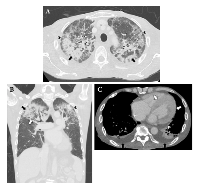
A 56-year-old male with underlying light chain multiple myeloma and systemic amyloidosis presented with fever for 2 days without dyspnea or cough. Further chest imaging revealed nonspecific findings including consolidations, ground-glass opacities, interlobular septal thickening in both upper lobes, and bilateral pleural effusions; a diagnosis of pneumonia with pulmonary edema was made. After the patient failed to respond to treatment, bronchoscopy with tissue biopsy was performed for unresolving pneumonia. Histopathological results were consistent with pulmonary amyloidosis.
Downloads
Metrics
References
Czeyda-Pommersheim F, Hwang M, Chen SS, Strollo D, Fuhrman C, Bhalla S. Amyloidosis: modern cross-sectional imaging. Radiographics 2015;35:1381–92. doi: 10.1148/rg.2015140179.
Baumann B, Salina D, Aboulhosn K. Clinical pulmonary amyloidosis presenting as lung cavitation with bronchiectasis: a case report. BCMJ 2019;61:344-8.
Blancas-Mejía LM, Ramirez-Alvarado M. Systemic amyloidoses. Annu Rev Biochem 2013;82:745–74. doi: 10.1146/annurev-biochem-072611-130030.
Li G, Han D, Wei S, Wang H, Chen L. Multiorgan involvement by amyloid light chain amyloidosis. J Int Med Res 2019;47:1778–86. doi: 10.1177/0300060518814337.
Lal A, Akhtar J, Khan MS, Chen Y, Yaron Goldman. Primary endobronchial amyloidosis: a rare case of endobronchial tumor. Respir Med Case Rep 2018;23:163–6. doi: 10.1016/j.rmcr.2018.02.007.
Georgiades CS, Neyman EG, Barish MA, Fishman EK. Amyloidosis: review and CT manifestations. Radiographics 2004;24:405–16. doi: 10.1148/rg.242035114.
Lee SH, Ko YC, Jeong JP, Park CW, Seo SH, Kim JT, et al. Single nodular pulmonary amyloidosis: case report. Tuberc Respir Dis (Seoul) 2015;78:385–9. doi: 10.4046/trd.2015.78.4.385.
Oda S, Kidoh M, Nagayama Y, Takashio S, Usuku H, Ueda M, et al. Trends in diagnostic imaging of cardiac amyloidosis: emerging knowledge and concepts. Radiographics 2020;40:961–81. doi: 10.1148/rg.2020190069.
Downloads
Published
How to Cite
Issue
Section
License
Copyright (c) 2020 The ASEAN Journal of Radiology

This work is licensed under a Creative Commons Attribution-NonCommercial-NoDerivatives 4.0 International License.
Disclosure Forms and Copyright Agreements
All authors listed on the manuscript must complete both the electronic copyright agreement. (in the case of acceptance)


