Calcific uraemic arteriolopathy: A rare but devastating complication of end-stage renal failure
DOI:
https://doi.org/10.46475/asean-jr.v25i2.897Keywords:
calcific uraemic arteriolopathy, calciphylaxis, renal failureAbstract
Calcific uraemic arteriolopathy is a rare complication of end-stage renal failure. It has a grave prognosis with 1-year survival of under 50%. It occurs due to subcutaneous small vessel calcification, thrombosis, with subsequent tissue necrosis.
Calcific uraemic arteriolopathy is a rare complication of end-stage renal failure. It carries a grave prognosis with 1-year survival of under 50%. It occurs due to subcutaneous small vessel calcification, thrombosis, with subsequent tissue necrosis.
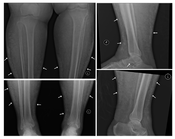
We described a case of calcific uraemic arteriolopathy in a 58-year-old man who presented with violaceous indurations over bilateral lower limbs, as well as large necrotic ulcer with adjacent eschars at the lower abdomen. Although skin biopsy is the gold standard for diagnosis, it is often avoided due to potential poor wound healing. On the other hand, in radiographs or computed tomography, fine linear or serpiginous subcutaneous calcifications are typical manifestations, which represent underlying small vessel calcifications. Radiological examinations, therefore, play an important role to establish the diagnosis.
Downloads
Metrics
References
Colboc H, Moguelet P, Bazin D, Carvalho P, Dillies AS, Chaby G, et al. Localization, morphologic features, and chemical composition of calciphylaxis-related skin deposits in patients with calcific uremic arteriolopathy. JAMA Dermatol 2019;155:789-96. doi: 10.1001/jamadermatol.2019.0381. DOI: https://doi.org/10.1001/jamadermatol.2019.0381
Weenig RH, Sewell LD, Davis MD, McCarthy JT, Pittelkow MR. Calciphylaxis: natural history, risk factor analysis, and outcome. J Am Acad Dermatol 2007;56:569-79. doi: 10.1016/j.jaad.2006.08.065. DOI: https://doi.org/10.1016/j.jaad.2006.08.065
Jovanovich A, Chonchol M. Calcific uremic arteriolopathy revisited. J Am Soc Nephrol 2016;27:3233-5. doi: 10.1681/ASN.2016040480. DOI: https://doi.org/10.1681/ASN.2016040480
Jeong HS, Dominguez AR. Calciphylaxis: controversies in pathogenesis, diagnosis and treatment. Am J Med Sci 2016;351:217-27. doi: 10.1016/j.amjms.2015.11.015. DOI: https://doi.org/10.1016/j.amjms.2015.11.015
Bleibel W, Hazar B, Herman R. A case report comparing various radiological tests in the diagnosis of calcific uremic arteriolopathy. Am J Kidney Dis 2006;48:659-61. doi: 10.1053/j.ajkd.2006.05.031. DOI: https://doi.org/10.1053/j.ajkd.2006.05.031

Downloads
Published
How to Cite
Issue
Section
License
Copyright (c) 2024 The ASEAN Journal of Radiology

This work is licensed under a Creative Commons Attribution-NonCommercial-NoDerivatives 4.0 International License.
Disclosure Forms and Copyright Agreements
All authors listed on the manuscript must complete both the electronic copyright agreement. (in the case of acceptance)


