Evaluation of the clinical utility of liver MRI T2* as a surrogate marker in liver iron overload: Experience from a Malaysian haematology referral centre
DOI:
https://doi.org/10.46475/asean-jr.v27i2.1017Keywords:
Ferritin, Iron, Liver iron concentration, Malaysia, MRI T2*, ThalassaemiaAbstract
Introduction: Thalassaemia significantly contributes to the disease burden in Southeast Asia, particularly due to complications such as liver iron overload from long-term blood transfusions. The assessment of liver iron overload moves from liver biopsy towards non-invasive methods by utilizing MRI T2* technique. This study evaluated the clinical utility of MRI T2* as a gold standard for assessing hepatic iron overload, and investigated its correlation with serum ferritin and other clinical parameters.
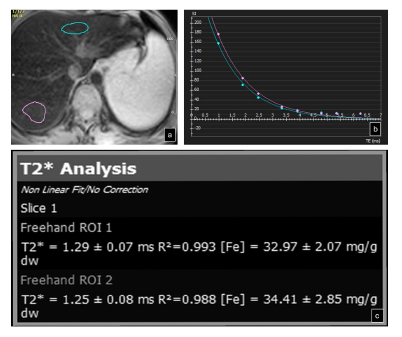
Materials and Methods: A one-year cross-sectional study was conducted on Thalassaemia patients who underwent MRI T2* in the year 2025. Liver iron concentration (LIC) was calculated using cvi-42 post-processing software and the serum ferritin levels, transfusion regimen, and demographic data were recorded. The relationships between T2* values, serum ferritin, and other clinical parameters were then analysed.
Results: A total of 158 Thalassaemia patients, predominantly transfusion-dependent, were enrolled. HbE-Beta Thalassaemia was the most common subtype (48.1%), followed by Beta-Thalassaemia Major (24.7%). Severe liver iron overload was observed in 63.3% of patients, with Malay patients exhibiting higher severity than Chinese patients (p<0.001). Serum ferritin levels showed a strong negative correlation with liver T2* values (Rs = -0.699, p < 0.001)However, the type of Thalassaemia, transfusion duration and regimen were not significantly associated with T2* values or LIC.
Conclusion: Liver MRI T2* demonstrated an effective assessment of hepatic iron overload, exhibiting a strong correlation with serum ferritin levels. These findings elucidate the reliability of MRI T2* imaging to be utilized in the management of transfusion-related iron overload.
Downloads
Metrics
References
Modell B, Darlison M. Global epidemiology of haemoglobin disorders and derived service indicators. Bull World Health Organ 2008;86:480-7. doi: 10.2471/blt.06.036673. DOI: https://doi.org/10.2471/BLT.06.036673
Argyropoulou MI, Astrakas L. MRI evaluation of tissue iron burden in patients with beta-thalassaemia major. Pediatr Radiol 2007;37:1191-200; quiz 1308-9. doi: 10.1007/s00247-007-0567-1. DOI: https://doi.org/10.1007/s00247-007-0567-1
Weatherall DJ. The evolving spectrum of the epidemiology of thalassemia. Hematol Oncol Clin North Am 2018;32:165-75. doi: 10.1016/j.hoc.2017.11.008. DOI: https://doi.org/10.1016/j.hoc.2017.11.008
De Sanctis V, Kattamis C, Canatan D, Soliman AT, Elsedfy H, Karimi M, et al. β-Thalassemia distribution in the Old World: an ancient disease seen from a historical standpoint. Mediterr J Hematol Infect Dis 2017;9:e2017018. doi: 10.4084/MJHID.2017.018. DOI: https://doi.org/10.4084/mjhid.2017.018
Mohd Ibrahim H, Alias H, Muda Z, Gunasagaran K, Osman R. Annual report of the Malaysian Thalassaemia Registry 2019. Kuala Lumpur: Ministry of Health Malaysia; 2019.
Mishra AK, Tiwari A. Iron overload in beta-thalassaemia major and intermedia patients. Maedica (Bucur) 2013;8:328-32.
Taksande A, Prabhu S, Venkatesh S. Cardiovascular aspect of beta-thalassaemia. Cardiovasc Hematol Agents Med Chem 2012;10:25-30. doi: 10.2174/187152512799201172. DOI: https://doi.org/10.2174/187152512799201172
Coates TD. Physiology and pathophysiology of iron in hemoglobin-associated diseases. Free Radic Biol Med 2014;72:23-40. doi: 10.1016/j.freeradbiomed.2014.03.039. DOI: https://doi.org/10.1016/j.freeradbiomed.2014.03.039
Ministry of Health Malaysia. Clinical practice guidelines: Management of thalassaemia. 2nd ed. Kuala Lumpur: Ministry of Health Malaysia; 2024.
Taher AT, Saliba AN. Iron overload in thalassemia: different organs at different rates. Hematology Am Soc Hematol Educ Program 2017;2017:265-71. doi: 10.1182/asheducation-2017.1.265. DOI: https://doi.org/10.1182/asheducation-2017.1.265
St Pierre TG, Clark PR, Chua-anusorn W, Fleming AJ, Jeffrey GP, Olynyk JK, et al. Noninvasive measurement and imaging of liver iron concentrations using proton magnetic resonance. Blood 2005;105:855-61. doi: 10.1182/blood-2004-01-0177. DOI: https://doi.org/10.1182/blood-2004-01-0177
Kwiatkowski JL, Kim HY, Thompson AA, Quinn CT, Mueller BU, Odame I, et al. Chelation use and iron burden in North American and British thalassemia patients: a report from the Thalassemia Longitudinal Cohort. Blood 2012;119:2746-53. doi: 10.1182/blood-2011-04-344507. DOI: https://doi.org/10.1182/blood-2011-04-344507
Hernando D, Zhao R, Yuan Q, Aliyari Ghasabeh M, Ruschke S, Miao X, et al. Multicenter reproducibility of liver iron quantification with 1.5-T and 3.0-T MRI. Radiology 2023;306:e213256. doi: 10.1148/radiol.213256. DOI: https://doi.org/10.1148/radiol.213256
Tuo Y, Li Y, Li Y, Ma J, Yang X, Wu S, et al. Global, regional, and national burden of thalassemia, 1990-2021: a systematic analysis for the Global Burden of Disease Study 2021. EClinicalMedicine 2024;72:102619. doi: 10.1016/j.eclinm.2024.102619. DOI: https://doi.org/10.1016/j.eclinm.2024.102619
Hoe HG, Git KA, Loh CK, Abdul Latiff Z, Hong J, Abdul Hamid H, et al. Magnetic resonance imaging T2* of the pancreas value using an online software tool and correlate with T2* value of myocardium and liver among patients with transfusion-dependent thalassemia major. Front Radiol 2022;2:943102. doi: 10.3389/fradi.2022.943102. DOI: https://doi.org/10.3389/fradi.2022.943102
Khadivi Heris H, Nejati B, Rezazadeh K, Sate H, Dolatkhah R, Ghoreishi Z, et al. Evaluation of iron overload by cardiac and liver T2* in β-thalassemia: correlation with serum ferritin, heart function and liver enzymes. J Cardiovasc Thorac Res 2021;13:54-60. doi: 10.34172/jcvtr.2021.18. DOI: https://doi.org/10.34172/jcvtr.2021.18
Alwi ZB, Syed-Hassan SR. Thalassemia in Malaysia. Hemoglobin 2022;46:45-52. doi: 10.1080/03630269.2022.2057326. DOI: https://doi.org/10.1080/03630269.2022.2057326
Tan JA, Chin PS, Wong YC, Tan KL, Chan LL, George E. Characterisation and confirmation of rare beta-thalassaemia mutations in the Malay, Chinese and Indian ethnic groups in Malaysia. Pathology 2006;38:437-41. doi: 10.1080/00313020600922538. DOI: https://doi.org/10.1080/00313020600922538
Di Tucci AA, Matta G, Deplano S, Gabbas A, Depau C, Derudas D, et al. Myocardial iron overload assessment by T2* magnetic resonance imaging in adult transfusion-dependent patients with acquired anemias. Haematologica 2008;93: 1385-8. doi: 10.3324/haematol.12759. DOI: https://doi.org/10.3324/haematol.12759
Chaosuwannakit N, Makarawate P. The value of magnetic resonance imaging in evaluation of myocardial and liver iron overload in a thalassaemia endemic population: a report from Northeastern Thailand. Pol J Radiol 2019;84:e262-8. doi: 10.5114/pjr.2019.86094. DOI: https://doi.org/10.5114/pjr.2019.86094
Anderson LJ. Assessment of iron overload with T2* magnetic resonanceimaging. Prog Cardiovasc Dis 2011;54:287-94. doi: 10.5114/pjr.2019.86094. DOI: https://doi.org/10.1016/j.pcad.2011.07.004
Wood JC, Enriquez C, Ghugre N, Tyzka JM, Carson S, Nelson MD, et al. MRI R2 and R2* mapping accurately estimates hepatic iron concentration in transfusion-dependent thalassemia and sickle cell disease patients. Blood 2005;106:1460-5. doi: 10.1182/blood-2004-10-3982. DOI: https://doi.org/10.1182/blood-2004-10-3982
Downloads
Published
How to Cite
Issue
Section
License
Copyright (c) 2026 The ASEAN Journal of Radiology

This work is licensed under a Creative Commons Attribution-NonCommercial-NoDerivatives 4.0 International License.
Disclosure Forms and Copyright Agreements
All authors listed on the manuscript must complete both the electronic copyright agreement. (in the case of acceptance)