
Utility of screening chest radiographs in patients with asymptomatic and mildly symptomatic COVID-19 at a field hospital in Samut Sakhon, Thailand
DOI:
https://doi.org/10.46475/aseanjr.v22i2.119Keywords:
Covid-19, Chest radiographs, Field hospitalAbstract
Background: In a new episode of the COVID -19 pandemic in Thailand during the beginning of 2021, cases in Samut Sakhon Province mainly occurred in foreign workers and were mostly asymptomatic or had mild disease. To prevent overwhelming the local hospital, a field hospital was established which used chest radiography as one of screening tools for triaging patients.
Objective: To determine the clinical utility of chest radiographs as a screening tool for COVID-19 patients who were asymptomatic or mildly symptomatic.
Materials and Methods: Six hundred nineteen patients with COVID -19 (confirmed by reverse transcriptase-polymerase chain reaction) were registered at the field hospital at Samut Sakhon provincial sport stadium during 5-8 January 2021 and had chest radiographs taken. The image readings were based on the consensus of two radiologists and a final decision was made by a third radiologist if the first two did not agree. Findings on chest radiographs and clinical outcomes were evaluated.
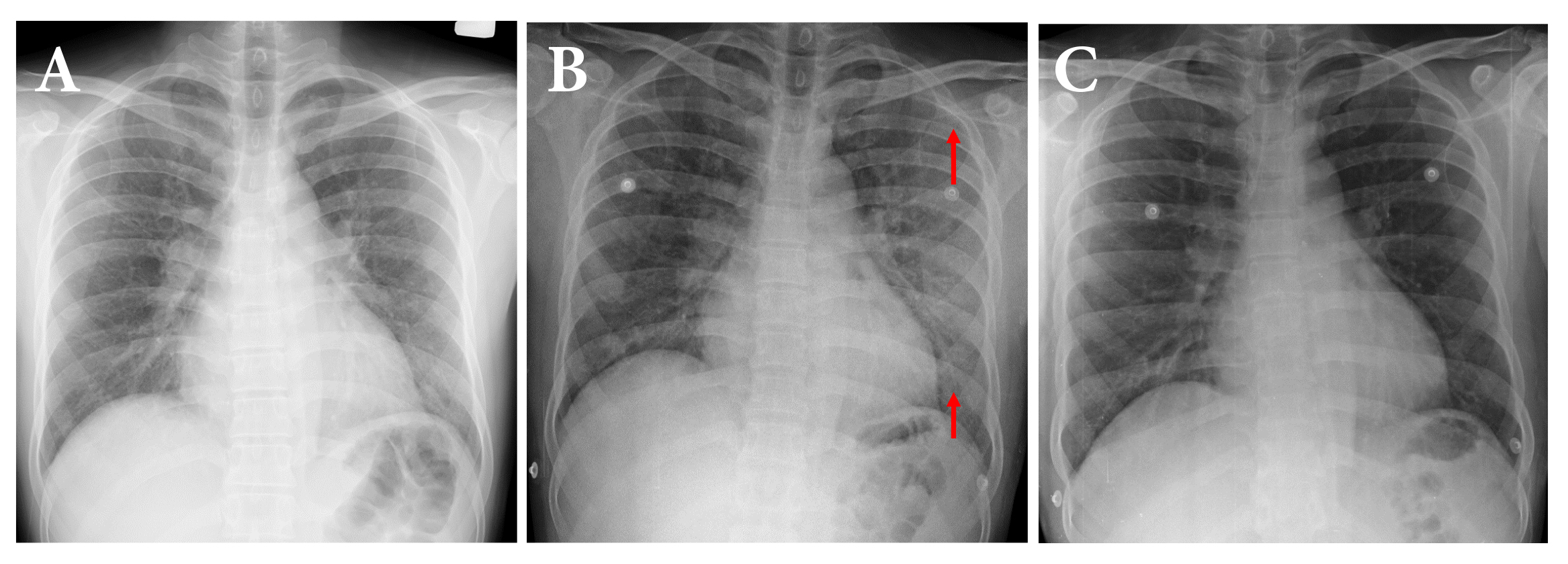
Results: The study included 619 radiographs; 328/619 (53%) men and 291/619 (47%) women had a mean age of 33.3+/- 9.7 (range, 5-64) years. There was mild disease in 13/619, and asymptomatic infections in 606/619. Chest radiographs were normal in 568 (91.7%) and abnormal in 51 (8.3%) patients; typical findings of COVID-19 were seen in 3 (0.5%) patients. Other abnormal findings were found in 23 (3.8%) patients such as active tuberculosis in 6 (1.0%). Four patients were transferred to the hospital, one of whom required supplemental oxygen.
Conclusion: Combined chest radiographic and clinical information allows better decisions regarding hospital transfers of asymptomatic and mildly symptomatic COVID-19 patients at a field hospital.
Downloads
Metrics
References
Huang C, Wang Y, Li X, Ren L, Zhao J, Hu Y, et al. Clinical features of patients infected with 2019 novel coronavirus in Wuhan, China. Lancet 2020;395:497-506. doi: 10.1016/S0140-6736(20)30183-5.
World Health Organization [Internet].Geneva: The Organization; 2021 [cited 2021 Jul 1]. Coronavirus Disease 2019 (COVID-19) Dashboard; [about 7 screens]. Available from: https:// www. Covidi2.who.int
Wu Z, Mcgoogan GM. Characteristics of and important lessons from the Coronavirus Disease 2019 (COVID-19) outbreak in China: summary of a report of 72314 cases from the Chinese Center for Disease Control and Prevention. JAMA 2020;323:1239-42. doi: 10.1001/jama.2020.2648.
Oran DP, Topol EJ. Prevalence of asymptomatic SARS-CoV-2 infection. Ann InternMed 2020;173:362-7. doi: 10.7326/M20-3012.
Li Q, Guan X, Wu P, Wang X, Zhou L, Tong Y, et al. Early transmission dynamics in Wuhan, China, of Novel Coronavirus-I-infected pneumonia. N Engl J Med 2020;382:1199-207. doi: 10.1056/NEJMoa2001316.
Guan WJ, Ni ZY, Hu Y, Liang WH, Ou CQ, He JX, et al. Clinical characteristics of Coronavirus Disease 2019 in China. N Engl J Med 2020;382:1708-20. doi: 10.1056/NEJMoa2002032.
Chinese Society of Radiology, Chinese Medical Association. Radiological diagnosis of New Corona Virus infected pneumonitis: expert recommendation from Chinese Society of Radiology (First Edition). Chin J Radiol [Internet] 2020 [cited 2021 Jul 1];4:279-85. Chinese. Available from: http://rs.yiigle.com/CN112149202004/1187937.htm
Zu ZY, Jiang MD, Xu PP, Chen W, Ni QQ, Lu GM, et al. Coronavirus Disease 2019 (COVID-19): a perspective from China. Radiology 2020;296:E15-25. doi: 10.1148/radiol.2020200490.
Ng MY, Lee EYP, Yang J, Yang F, Li X, Wang H, et al. Imaging profile of the COVID-19 infection: radiologic findings and literature review. Radiol Cardiothorac Imaging 2020;2:e200034. doi: 10.1148/ryct.2020200034.
Stephanie S, Shum ST, Cleveland H, Challa SR, Herring A, Jacobson FL,et al. Determinants of chest X-Ray sensitivity for COVID-19: a multi-institutional study in the United States. Radiol Cardiothorac Imaging 2020;2: e200337. doi: 10.1148/ryct.2020200337
Rubin GD, Ryerson CJ, Haramati LB, Sverzellati N, Kanne JP, Raoof S, et al. The role of chest imaging in patient management during the COVID-19 pandemic: a multinational consensus statement from the Fleischner Society. Radiology 2020;296:172-80. doi: 10.1148/radiol.2020201365.
ACR American College of Radiology [Internet]. Reston(VA): ACR; 2020 Mar 11 [updated 2020 Mar 22; cited 2021 Jul 1]. ACR recommendations for the use of chest radiography and Computed Tomography (CT) for suspected COVID-19 infection. Available from: https://www.acr.org/Advocacy-and-Economics/ACR-Position-Statements/Recommendations-for-Chest-Radiography-and-CT-for-Suspected-COVID19-Infection.
World Health Organization [Internet]. Geneva: WHO; 2020 Apr 2 [cited 2021 Jul 1]. Coronavirus Disease 2019 (COVID-19) situation report -73. Available from: https://www.who.int/docs/default-source/coronaviruse/situation-reports/20200402-sitrep-73-covid-19.pdf?sfvrsn=5ae25bc7_6.
World Health Organization [Internet]. Geneva: WHO; 2020 [cited 2021 Jul 1].Use of chest imaging in COVID-19: a rapid advice guide 11 June 2020. Available from: https://apps.who.int/iris/handle/10665/332336
Litmanovich DE, Chung M, Kirkbride RR,, Kicska G, Kanne JP. Review of chest radiograph findings of COVID-19 pneumonia and suggested reporting language. J Thorac Imaging 2020;35:354-60. doi: 10.1097/RTI.0000000000000541.
Borghesi A, Maroldi R. COVID-19 outbreak in Italy: experimental chest X-ray scoring system for quantifying and monitoring disease progression. Radiol Med 2020;125:509-13. doi: 10.1007/s11547-020-01200-3.
Hui TCH, Khoo HW, Young BE, Haja Mohideen SM, Lee YS, Lim CJ, et al. Clinical utility of chest radiography for severe COVID-19. Quant Imaging Med Surg 2020;10:1540-50. doi: 10.21037/qims-20-642.
Kuo BJ, Lai YK, Tan MLM, Goh XC. Utility of screening chest radiographs in patients with asymptomatic or minimally symptomatic COVID-19 in Singapore. Radiology 2021;298:E131-40. doi: 10.1148/radiol.2020203496.
Parry AH, Wani AH, Yaseen M, Shah NN, Dar KA. Clinicoradiological course in coronavirus disease-19 (COVID-19) patients who are asymptomatic at admission. BJR Open 2020;2:20200033.doi: 10.1259/bjro.20200033.
Wong HYF, Lam HYS, Fong AH, Leung ST, Chin TW, Lo CSY, et al. Frequency and distribution of chest radiographic findings in patients positive for COVID-19. Radiology. 2020;296:E72-8. doi: 10.1148/radiol.2020201160.
Downloads
Published
How to Cite
Issue
Section
License
Copyright (c) 2021 The ASEAN Journal of Radiology

This work is licensed under a Creative Commons Attribution-NonCommercial-NoDerivatives 4.0 International License.
Disclosure Forms and Copyright Agreements
All authors listed on the manuscript must complete both the electronic copyright agreement. (in the case of acceptance)


