Imaging Findings of OHVIRA Syndrome with Surgical Correlation in an 11-Year-Old Girl: A classic case
DOI:
https://doi.org/10.46475/asean-jr.v27i2.1021Keywords:
OHVIRA syndrome, Obstructed hemivagina, Renal agenesis, Ultrasound, Computed tomographyAbstract
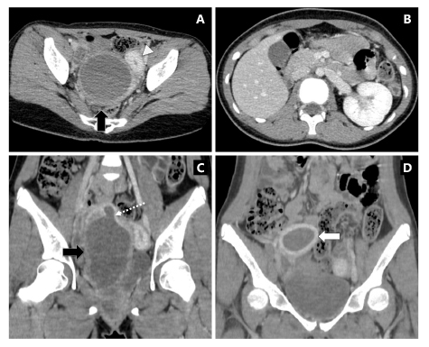
OHVIRA syndrome (Obstructed Hemivagina and Ipsilateral Renal Agenesis) is a rare congenital Müllerian duct anomaly characterized by uterine duplication, obstructed hemivagina, and ipsilateral renal agenesis. Early recognition is essential to prevent long-term complications such as endometriosis and infertility. We report the case of an 11-year-old girl who presented with acute lower abdominal pain for one day without fever. Physical examination revealed a 10-cm vaginal mass. Transabdominal ultrasonography demonstrated a cystic mass in the mid-pelvic cavity. Contrast-enhanced computed tomography confirmed uterine didelphys with marked distension of the right hemivagina and absence of the right kidney. Surgical exploration revealed a bulging right hemivagina with approximately 400 mL of retained brownish fluid, and vaginal septum excision was performed. The postoperative course was uneventful. This case highlights the importance of multimodality imaging in the early diagnosis and management of OHVIRA syndrome.
Downloads
Metrics
References
Taliento C, Rondoni A, Scutiero G, Pavone M, Mordenti M, Vizzieli G, et al. Uterus didelphys with hematocervix in the unilateral obstructed hemivagina and ipsilateral renal agenesis: a case report of Herlyn-Werner-Wunderlich syndrome. Reprod Med [Internet]. 2024 [cited 2026 Mar 28];5:197-203. Available from: https://doi.org/10.3390/reprodmed5040018 DOI: https://doi.org/10.3390/reprodmed5040018
Burgis J. Obstructive Müllerian anomalies: case report, diagnosis, and management. Am J Obstet Gynecol 2001;185:338–44. doi: 10.1067/mob.2001.116738. DOI: https://doi.org/10.1067/mob.2001.116738
Devanabanda B, Chatterton B, Hassan IN, Patel J. Obstructed hemivagina with ipsilateral renal agenesis (OHVIRA) syndrome: typical presentation of a rare syndrome. Radiol Case Rep 2023;18:2437-40. doi: 10.1016/j.radcr.2023.04.016. DOI: https://doi.org/10.1016/j.radcr.2023.04.016
Del Vescovo RD, Battisti S, Di Paola VD, Piccolo CL, Cazzato RL, Sansoni I, et al. Herlyn-Werner-Wunderlich syndrome: MRI findings, radiological guide (two cases and literature review), and differential diagnosis. BMC Med Imaging 2012;12:4. doi: 10.1186/1471-2342-12-4. DOI: https://doi.org/10.1186/1471-2342-12-4
Behr SC, Courtier JL, Qayyum A. Imaging ofMüllerian duct anomalies. Radiographics 2012;32:E233-50. doi: 10.1148/rg.326125515. DOI: https://doi.org/10.1148/rg.326125515
Kim HJ, Lee EJ, Chang YW, Hong SS, Hwang J, Nam B, et al. Anatomical variations, genitourinary anomalies and clinical presentations in obstructed hemivagina and ipsilateral renal anomaly syndrome: case series. J Korean Soc Radiol 2025;86:129-40. doi: 10.3348/jksr.2024.0018. DOI: https://doi.org/10.3348/jksr.2024.0018
Zhu L, Chen N, Tong JL, Wang W, Zhang L, Lang JH. New classification of Herlyn‑Werner‑Wunderlich syndrome. Chin Med J (Engl) 2015;128:222-5. doi: 10.4103/0366-6999. DOI: https://doi.org/10.4103/0366-6999.149208
Tong J, Zhu L, Chen N, Lang J. Endometriosis in association with Herlyn-Werner-Wunderlich syndrome. Fertil Steril 2014;102:790-4. doi: 10.1016/j.fertnstert.2014.05.025. DOI: https://doi.org/10.1016/j.fertnstert.2014.05.025
Smith NA, Laufer MR. Obstructed hemivagina and ipsilateral renal anomaly
(OHVIRA) syndrome: management and follow-up. Fertil Steril 2007;87:918-22. doi: 10.1016/j.fertnstert.2006.11.015. DOI: https://doi.org/10.1016/j.fertnstert.2006.11.015
Downloads
Published
How to Cite
Issue
Section
License
Copyright (c) 2026 The ASEAN Journal of Radiology

This work is licensed under a Creative Commons Attribution-NonCommercial-NoDerivatives 4.0 International License.
Disclosure Forms and Copyright Agreements
All authors listed on the manuscript must complete both the electronic copyright agreement. (in the case of acceptance)