
Quantitative CT characterization of normal lung anatomy: In relation to locations and gender and study of various lung volume estimation
Keywords:
Mean lung attenuation, Lung volume estimation, Ellipsoid formulaAbstract
Background: Mean lung parenchymal attenuation (MLA) may show gender differences and vary in different locations. Lung volume estimations play an important role in lung transplantation workout. In the current study, we focus on quantitative measurement of lung volume using different estimations obtained through calculated formulae from CT images and Chest Radiograph. This study could help to generate data for future references, particularly for Malaysia or Southeast Asia.
Objective: To study the MLA in relation to the location, patient’s position and gender differences, in order to find the correlation of various lung volume estimation methods.
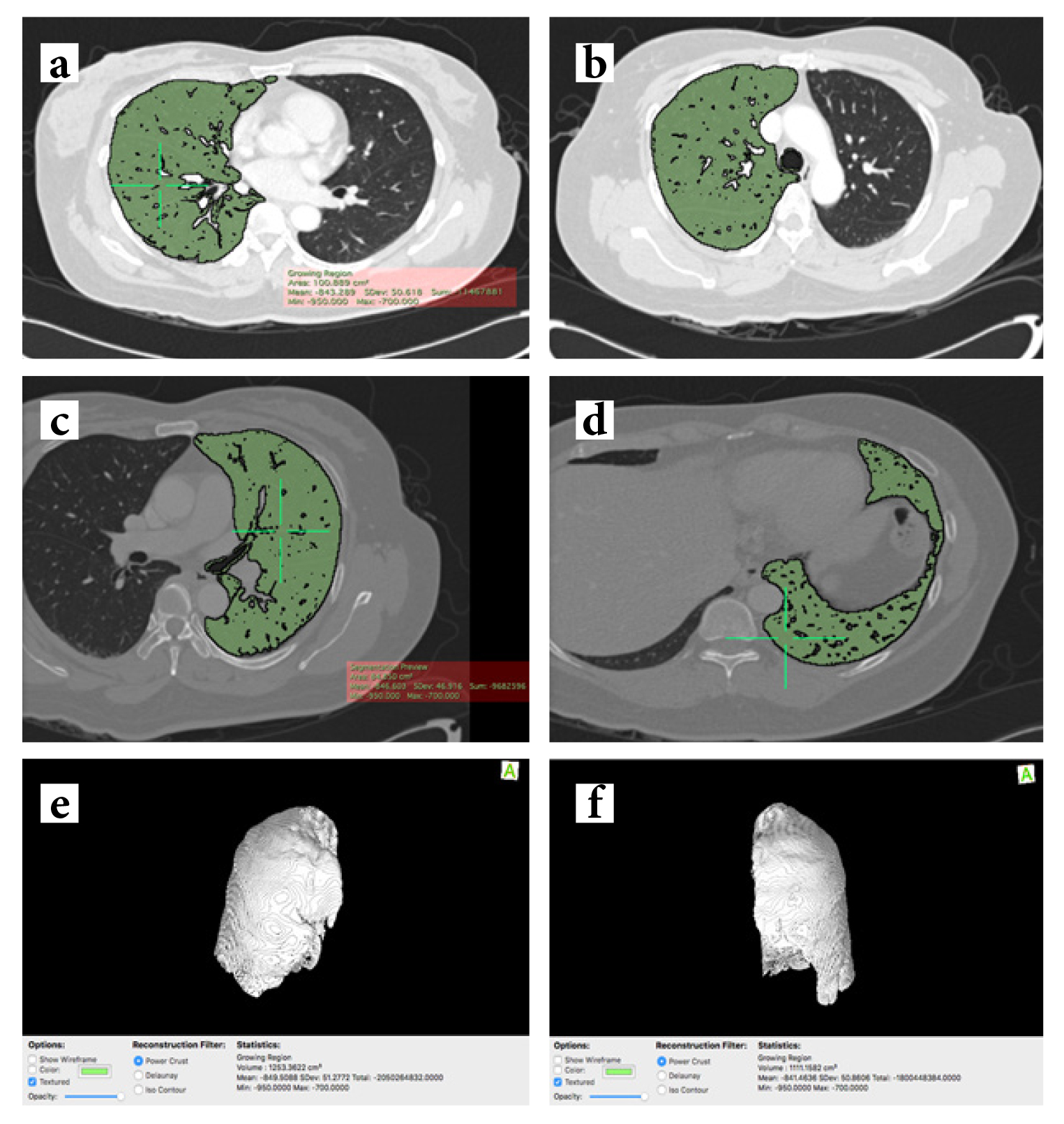
Materials and Methods: This was a retrospective cross-sectional study. A total of 62 patients’ CT images were selected and measurements of MLA were analyzed at lung apex, main carinal level (MCL), and base level (BL). At MCL and BL, the MLA was measured in three regions which were anterior, middle and posterior regions. A total lung volume of 26 from the 62 study subjects was also estimated using the predictive equation (method i), estimation from the frontal chest radiograph (method ii), ellipsoid formula using measurement from CT images (method iii) and semi auto-segmentation and volumetric calculation (method iv).
Results: MLA of the right lungs range from -860±9.52 to -787.66±14.8 HU. MLA of the left lungs range from -845.60±10.0 to -789.66±14.0 HU. MLA at middle MCL is lower than apex and middle BL bilaterally (p<0.05). Male subjects have lower lung attenuation with several areas showing a statistically significant difference (p<0.05). The four lung volume estimations show moderate to strong linear correlation (r = 0.565 – 0.899).
Conclusion: Variation of MLA among MCL, apex and BL could be related to better chest expansion at MCL. Theposterior part of the lungs shows a higher value of MLA and this is related to the supine position during scanning that results in a lesser degree of chest expansion at the posterior part and gravity influence. Lung volume estimation is possible using demographic data, a chest radiograph or the ellipsoid formula.
Downloads
Metrics
References
Whiting P, Singatullina N, Rosser JH. Computed tomography of the chest: I. Basic principles. BJA Educ 2015;15:299-304. doi:10.1093/bjaceaccp/mku063.
Haas M, Hamm B, Niehues SM. Automated lung volumetry from routine thoracic CT scans: how reliable is the result? Acad Radiol 2014;21:633-8. doi: 10.1016/j.acra.2014.01.002.
Stein JM, Walkup LL, Brody AS, Fleck RJ, Woods JC. Quantitative CT characterization of pediatric lung development using routine clinical imaging. Pediatr Radiol 2016;46:1804-12. doi: 10.1007/s00247-016-3686-8.
Zach JA, Newell JD Jr, Schroeder J, Murphy JR, Curran-Everett D, Hoffman EA, et al. Quantitative computed tomography of the lungs and airways in healthy nonsmoking adults. Invest Radiol 2012;47:596-602. doi: 10.1097/RLI.0b013e318262292e.
Smit HJ, Golding RP, Schramel FM, Devillé WL, Manoliu RA, Postmus PE. Lung attenuation measurements in healthy young adults. Respiration 2003;70:143-8. doi: 10.1159/000070060.
Kauczor HU, Hast J, Heussel CP, Schlegel J, Mildenberger P, Thelen M. CT attenuation of paired HRCT scans obtained at full inspiratory/expiratory position: comparison with pulmonary function tests. Eur Radiol 2002;12:2757-63. doi: 10.1007/s00330-002-1514-z.
Schlesinger AE, White DK, Mallory GB, Hildeboldt CF, Huddleston CB. Estimation of total lung capacity from chest radiography and chest CT in children: comparison with body plethysmography. AJR Am J Roentgenol 1995;165:151-4. doi: 10.2214/ajr.165.1.7785574.
Reger RB, Young A, Morgan WK. An accurate and rapid radiographic method of determining total lung capacity. Thorax 1972;27:163-8. doi: 10.1136/thx.27.2.163.
Canals M, Olivares R, Rosenmann M. A radiographic method to estimate lung volume and its use in small mammals. Biol Res 2005;38:41-7. doi: 10.4067/s0716-97602005000100006.
Konheim JA, Kon ZN, Pasrija C, Luo Q, Sanchez PG, Garcia JP, et al. Predictive equations for lung volumes from computed tomography for size matching in pulmonary transplantation. J Thorac Cardiovasc Surg 2016;151:1163-9. doi: 10.1016/j.jtcvs.2015.10.051.
Cheng T, Li Y, Pang S, Wan H, Shi G, Cheng Q, et al. Normal lung attenuation distribution and lung volume on computed tomography in a Chinese population. Int J Chron Obstruct Pulmon Dis 2019;14:1657-68. doi: 10.2147/COPD.S187596.
Lee KN, Yoon SK, Sohn CH, Choi PJ, Webb WR. Dependent lung opacity at thin-section CT: evaluation by spirometrically-gated CT of the influence of lung volume. Korean J Radiol 2002;3:24-9. doi: 10.3348/kjr.2002.3.1.24.
Kim SS, Jin GY, Li YZ, Lee JE, Shin HS. CT quantification of lungs and airways in normal Korean subjects. Korean J Radiol 2017;18:739-48. doi:10.3348/kjr.2017.18.4.739.
Adedoyin R, Adeleke OE, Fehintola AO, Erhabor G, Bisiriyu L. Reference values for chest expansion among adult residents in Ile-Ife, Nigeria- a cross-sectional study. J Phys Ther 2013;6:54-8. doi:10.4172/2157-7595.1000113.
LoMauro A, Aliverti A. Sex differences in respiratory function. Breathe (Sheff) 2018;14:131-40. doi:10.1183/20734735.000318.
Akoglu H. User's guide to correlation coefficients. Turk J Emerg Med 2018;18:91-3. doi: 10.1016/j.tjem.2018.08.001.
Kovacevic M, Goranovic T, Markic A, Jelisavac M, Zuric I, Tonkovic D. Usefulness of routine chest X-ray in preoperative evaluation of patients undergoing non-cardiopulmonary surgery: a prospective observational study:1AP5-5 [abstract]. Eur J Anaesthesiol 2012;29 Suppl 50:16.
Young EM, Farmer JD. Preoperative chest radiography in elective surgery: review and update. S D Med 2017;70:81-7.
Downloads
Published
How to Cite
Issue
Section
License
Copyright (c) 2021 The ASEAN Journal of Radiology

This work is licensed under a Creative Commons Attribution-NonCommercial-NoDerivatives 4.0 International License.
Disclosure Forms and Copyright Agreements
All authors listed on the manuscript must complete both the electronic copyright agreement. (in the case of acceptance)


