
Comparison of entrance surface air kerma measurement with MTS-N (LiF: Mg, Ti) chips with a kilovoltage X-ray source
DOI:
https://doi.org/10.46475/aseanjr.v22i1.96Keywords:
Entrance surface air kerma, Backscatter Radiation, Accuracy, Ionization Chamber, Detector, MTS-N ChipsAbstract
Objective: Radiation detectors are key components that ensure the accuracy and performances of dosimetry equipment. The study is aimed to compare the mean entrance surface air kerma (ESAK) between a DCT-10mm ionization chamber (IC) and MTS-N (LiF: Mg, Ti) chips when both detectors are exposed to ≤ 5mGy with a 10 by 10 field size, with an X-ray source and to determine the accuracy of the Thermoluminescent (TL) chips. Also, the dose will be compared to similar studies.
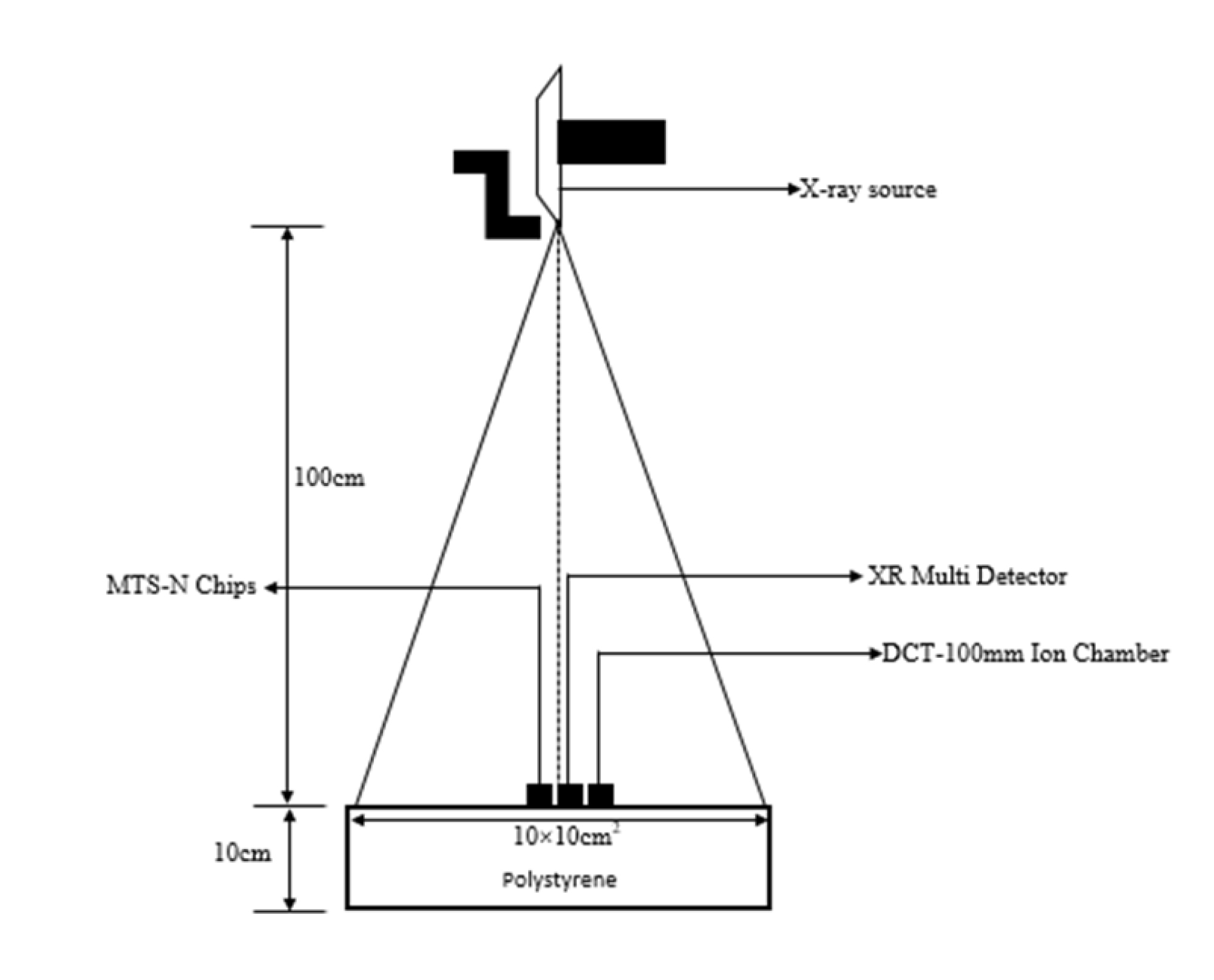
Materials and Methods: A functional, Digital Radiography (DR) X-ray System was used. A DCT-10mm ionization chamber (IC) and an XR Multidetector was positioned at a Source to Image Distance (SID) of 100cm on polystyrene, about 20cm thick. An X-ray spectrum generated at a Practical Peak Voltage (PPV) of 60-107kV with Half Value Layer (HVL) of 2.4-4.3mmAl and filtration > 3mmAl was used. The same setup was used for the MTS-N chips.
Results: The mean doses for 1-5 mGy with the MTS-N chips were 1.07±0.07, 1.60±0.13, 2.23±0.11, 2.58±0.07 and 3.45±0.10 mGy respectively, with accuracies of 7, 20, 26, 36 and 31%. Dose accuracy at 1and 2mGy was within 25% respectively. Dose accuracies at 3, 4 and 5mGy was within >25%. The correction factor for 1-5mGy was 0.94, 1.25, 1.35, 1.55 and 1.45 respectively.
Conclusion: Validation of the MTS-N chips with the reference ionization chamber to this study was within 36%. The Radiation and Nuclear Safety Authority (STUK) recommends that ESAK be within 25% for entrance surface dose. ESAK accuracy mostly increased with dose as observed in this study.
Downloads
Metrics
References
Do KH. General principles of radiation protection in fields of diagnostic medical exposure. J Korean Med Sci 2016;31 Suppl 1 :S6-9. doi: 10.3346/jkms.2016.31.S1.S6.
Awosan KJ, Ibrahim M, Saidu SA, Ma'aji SM, Danfulani M, Yunusa EU, et al. Knowledge of radiation hazards, radiation protection practices and clinical profile of health workers in a teaching hospital in Northern Nigeria. J Clin Diagn Res 2016; 10(8): LC07-12. doi: 10.7860/JCDR/2016/20398.8394.
Mettler FA Jr, Thomadsen BR, Bhargavan M, Gilley DB, Gray JE, Lipoti JA, et al. Medical radiation exposure in the U.S. in 2006: preliminary results. Health Phys 2008; 95: 502-7. doi: 10.1097/01.HP.0000326333.42287.a2.
Sistrom CL, McKay NL. Costs, charges, and revenues for hospital diagnostic imaging procedures: differences by modality and hospital characteristics. J Am Coll Radiol 2005;2:511-9. doi: 10.1016/j.jacr.2004.09.013.
Bercovich E, Javitt MC. Medical imaging: from roentgen to the digital revolution, and beyond. Rambam Maimonides Med J 2018;9(4):e0034. doi: 10.5041/RMMJ.10355.
Howell JD. Early clinical use of the X-ray. Trans Am Clin Climatol Assoc 2016; 127:341-9.
Obed RI, Ekpo ME, Omojola AD, Abdulkadir MK. Medical physics professional development and education in Nigeria. Med Phys Int 2016;4:96-8.
International Atomic Energy Agency,Dosimetry in diagnostic radiology: an international code of practice. Vienna: IAEA; 2007. Technical Reports Series No.: 457.
Horowitz YS, Satinger D, Fuks E, Oster L, Podpalov L. On the use of LiF:Mg,Ti thermoluminescence dosimeters in space-a critical review. Radiat Prot Dosimetry 2003; 106:7-24. doi: 10.1093/oxfordjournals.rpd.a006337.
Sadeghi M, Sina S, Faghihi R. Investigation of LiF, Mg and Ti (TLD-100) reproducibility. J Biomed Phys Eng 2015;5:217-22.
Horowitz Y, Oster L, Eliyahu I. Review of dose-rate effects in the thermoluminescence of LiF: Mg, Ti (HARSHAW). Radiat Prot Dosimetry 2018;179:184-8. doi: 10.1093/rpd/ncx248.
Hasegawa H, Sato M, Tanaka H. Evaluation of an X-Ray dose profile derived from an optically stimulated luminescent dosimeter during Computed Tomographic Fluoroscopy. PLoS ONE 2015;10(7):e0132154. doi: 10.1371/journal.pone.0132154.
Choi Y, Shil Cha E, Jin Bang Y, Ko S, Ha M, Lee C, et al. Estimation of organ doses among diagnostic medical radiation workers in South Korea. Radiat Prot Dosimetry 2018;179:142-50. doi: 10.1093/rpd/ncx239.
Ivanauskaite D, Griciene B. Status of individual dosimetry for dentists in Lithuania in year 1996-2001. Stomatologija 2003;5:149-51.
Sharma J, Sarma J, Agarwal S. Assessment of diagnostic reference level in radiography of neonatal chest anteroposterior examination: a hospital-based study. J Med Phys 2018;43:200-3. doi: 10.4103/jmp.JMP_37_18.
Institute of Physical Sciences in Medicine. National protocol for patient dose measurement in diagnostic radiology. Chilton:NRPB;1992.
Raffi RA, Davis SD, Hammer CG, Micka JA, Kunugi KA, Musgrove JE; et al. Determination of exit skin dose for 192Ir intracavitary accelerated partial breast irradiation with thermoluminescent dosimeters. Med Phys 2010;37: 2693-702. doi: 10.1118/1.3429089.
Omojola AD, Akpochafor MO, Adeneye SO, Aweda MA. Calibration of MTS N (LiF: Mg, Ti) chips using cesium 137 source at low doses for personnel dosimetry in diagnostic radiology. Radiat Prot Environ 2020;43:108-14.
Radiation and Nuclear Safety Authority (STUK). Radiation practices and radiation measurements. Helsinki: STUK; 2016.
Nilsson B, Sorcini B. Surface dose measurements in clinical photon beams. Acta Oncol 1989;28:537-42. doi: 10.3109/02841868909092265.
Yusof MFM, Yahya MH, Rosnan MS, Abdullah R, Abdul Kadir AB. Dose measurement using Al2O3 dosimeter in comparison to LiF: Mg, Ti dosimeter and ionization chamber at low and high energy X-ray. AIP Conf Proc 2017;1799:040007.doi: 10.1063/1.4972931.
Herrati A, Bourouina M, Khalal-Kouache K. Investigation of TLD-700 energy response to low energy x-ray encountered in diagnostic radiology. Open Phys 2016;14:150-8. doi: 10.1515/phys-2016-0016.
Alashrah S, Kandaiya S, Maalej N, El-Taher A. Skin dose measurements using radiochromic films, TLDs and ionisation chamber and comparison with Monte Carlo simulation. Radiat Prot Dosimetry 2014;162:338-44. doi: 10.1093/rpd/nct315.
Reynolds TA, Higgins P. Surface dose measurements with commonly used detectors: a consistent thickness correction method. J Appl Clin Med Phys 2015;16:358-66. doi: 10.1120/jacmp.v16i5.5572.
Fitriandini A, Wibowo WE, Pawiro SA. Comparison of dosimeter response: ionization chamber, TLD, and Gafchromic EBT2 film in 3D-CRT, IMRT, and SBRT techniques for lung cancer. J Phys:Conf Ser 2016;694:012006. doi: 10.1088/1742-6596/694/1/012006.
Waqar M, Ul-Haq A, Bilal S, Mosood M. Comparison of dosimeter response of TLD-100 and ionization chamber for high energy photon beams at KIRAN Karachi in Pakistan. Egypt J Radiol Nucl Med 2017; 48 :479-83. doi:10.1016/j.ejrnm.2017.01.012
Lee JH, Chang LT, Shiau AC, Chen CW, Liao YJ, Li WJ,et al. A novel simple phantom for verifying the dose of radiation therapy. BioMed Res Int 2015 ;2015:934387. doi: 10.1155/2015/934387.
Downloads
Published
How to Cite
Issue
Section
License
Copyright (c) 2021 The ASEAN Journal of Radiology

This work is licensed under a Creative Commons Attribution-NonCommercial-NoDerivatives 4.0 International License.
Disclosure Forms and Copyright Agreements
All authors listed on the manuscript must complete both the electronic copyright agreement. (in the case of acceptance)


