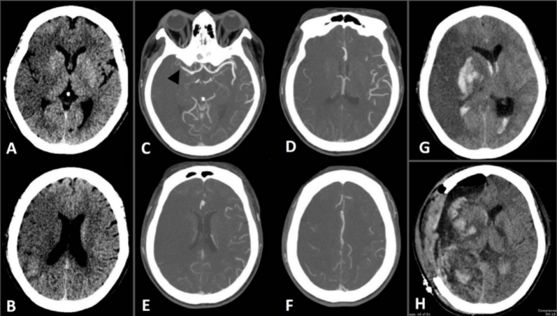
Value of computed tomography angiographic collateral status in prediction of malignant middle cerebral artery infarction
DOI:
https://doi.org/10.46475/aseanjr.v21i1.51Keywords:
CTA collateral status, Malignant MCA infarction, NECT ASPECTS, Acute ischemic stroke.Abstract
Background: Cerebral collateral circulation is necessary to maintain cerebral blood flow and penumbra when arterial insufficiency occurred. Only a few studies about collateral status on development of malignant middle cerebral artery infarction (mMCAi) have been documented.
Objective: To determine whether collateral status evaluated by single phase computed tomographic angiography (CTA) help prediction of mMCAi in patients with large arterial occlusion whom not received endovascular treatment.
Material and Methods: We retrospectively reviewed patients with acute ischemic stroke in anterior circulation in our institute during January 2015 to December 2015. We analyzed clinical data, baseline National Institutes of Health Stroke Scale (NIHSS), Alberta Stroke Program Early CT Score (ASPECTS) on baseline nonenhanced computed tomography of the brain (NECT brain), and CTA collateral status. Malignant MCA infarction was defined according to clinical criteria.
Results: Thirty-five patients were included. Mean age was 68.8±15.56 years. Mean baseline NIHSS and baseline ASPECTS were 17(±5) and 6(±3), respectively. All patients received intravenous thrombolysis. CTA collateral status and baseline NECT ASPECTS significantly correlated with development of mMCAi (P-value = 0.007 and 0.001). Only baseline NECT ASPECTS was an independent predictive factor for mMCAi (OR 0.63, 95%CI 0.46-0.86, P-value =0.004). Patients with baseline NECT ASPECTS ? 7 were more likely develop mMCAi (OR 14.29 95%CI 1.57-129.94, P-value 0.018).
Conclusion: In acute stroke patients with proximal MCA or ICA occlusion received intravenous thrombolysis alone, baseline NECT ASPECTS and CTA collateral status were significantly correlate with development of mMCAi. However, only baseline ASPECTS ? 7 was an independent predictor for mMCAi.
Downloads
Metrics
References
Hacke W, Schwab S, Horn M, Spranger M, De Georgia M, von Kummer R. 'Malignant' middle cerebral artery territory infarction: clinical course and prognostic signs. Arch Neurol 1996;53:309-15.
Ropper AH, Shafran B. Brain edema after stroke. Clinical syndrome and intracranial pressure. Arch Neurol 1984;41:26-9.
Heiss WD. Malignant MCA infarction: pathophysiology and imaging for early diagnosis and management decisions. Cerebrovasc Dis 2016;41:1-7. https://doi.org/10.1159/000441627.
Kim H, Jin ST, Kim YW, Kim SR, Park IS, Jo KW. Predictors of malignant brain edema in middle cerebral artery infarction observed on CT angiography. J Clin Neurosci 2015; 22:554–60.
Subramaniam S, Hill MD. Massive cerebral infarction. Neurologist 2005;11:150-60.
Staykov D, Gupta R. Hemicraniectomy in malignant middle cerebral artery infarction. Stroke 2011;42:513-6. https://doi.org/10.1161/STROKEAHA.110.605642.
Flores A, Rubiera M, Ribó M, Pagola J, Rodriguez-Luna D, Muchada M, et al. Poor collateral circulation assessed by multiphase computed tomographic angiography predicts malignant middle cerebral artery evolution after reperfusion therapies. Stroke 2015;46:3149-53. https://doi.org/10.1161/STROKEAHA.115.010608.
Liebeskind DS. Collateral circulation. Stroke 2003;34:2279-84.

Downloads
Published
How to Cite
Issue
Section
License
Copyright (c) 2020 The ASEAN Journal of Radiology

This work is licensed under a Creative Commons Attribution-NonCommercial-NoDerivatives 4.0 International License.
Disclosure Forms and Copyright Agreements
All authors listed on the manuscript must complete both the electronic copyright agreement. (in the case of acceptance)


