Dual-energy spectral CT in assessment of pleural effusion
DOI:
https://doi.org/10.46475/aseanjr.v23i3.193Keywords:
Pleural effusion, Dual energy spectral computed tomography, Benign pleural effusion, Malignant pleural effusionAbstract
Background: Pleural effusion is a common clinical problem. The ability to differentiate between benign and malignant pleural effusions has many clinical benefits. However, few studies have investigated the diagnostic value of dual-energy spectral computed tomography (DECT) in pleural effusion.
Objective: To evaluate the diagnostic values of DECT for assessment of pleural effusion.
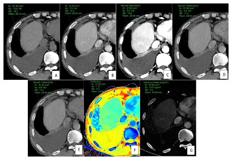
Materials and Methods: We retrospectively analyzed data from 87 patients presenting with pleural effusion who underwent chest DECT from October 2019 to November 2020. Two reviewers blindly reviewed the CT images of pleural effusion in consensus. The pleural fluid attenuation in standard conventional CT images and monoenergetic images, at 40 keV, 100 keV and 140 keV, were recorded. Data pertaining to the effective atomic number, the iodine concentration (IC)and associated CT findings including pleural thickening, pleural nodules and extrapleural fat clouding was analyzed.
Results: Of 87 patients, 44 were presented with benign effusions and the remaining 43 were presented with malignancy.There were no statistically significant differences in differentiating between benign and malignant pleural effusions by using the attenuation values, the effective atomic number or the IC. Irregular pleural thickening and pleural nodules were detected statistically significant in the patients with malignant pleural effusions with moderate accuracy, (48.83%; p < 0.01; AuROC 0.7103 and 34.88%; p < 0.01; AuROC 0.654 respectively).
Conclusion: DECT attenuation values did not show any reliable clinical value in the differentiation between benign and malignant pleural effusions. The presence of pleural nodules or irregular pleural thickening would suggest malignant pleural effusion with moderate accuracy.
Downloads
Metrics
References
Abramowitz Y, Simanovsky N, Goldstein MS, Hiller N. Pleural effusion: characterization with CT attenuation values and CT appearance. AJR Am J Roentgenol 2009;192:618-23. doi: 10.2214/AJR.08.1286.
Cullu N, Kalemci S, Karakas O, Eser I, Yalcin F, Boyaci FN, et al. Efficacy of CT in diagnosis of transudates and exudates in patients with pleural effusion. Diagn Interv Radiol 2014;20:116-20. doi: 10.5152/dir.2013.13066.
Zhang X, Duan H, Yu Y, Ma C, Ren Z, Lei Y, et al. Differential diagnosis between benign and malignant pleural effusion with dual-energy spectral CT. PLoS One 2018;13:e0193714. doi: 10.1371/journal.pone.0193714.
Porcel JM, Pardina M, Bielsa S, González A, Light RW. Derivation and validation of a CT scan scoring system for discriminating malignant from benign pleural effusions. Chest2015;147:513-9. doi: 10.1378/chest.14-0013.
Basso SMM, Lumachi F, Del Conte A, Sulfaro S, Maffeis F, Ubiali P. Diagnosis of Malignant Pleural Effusion Using CT Scan and Pleural-Fluid Cytology Together. A Preliminary Case-Control Study. Anticancer Res 2020;40:1135-9. doi: 10.21873/anticanres.14054.
Silva AC, Morse BG, Hara AK, Paden RG, Hongo N, Pavlicek W. Dual-energy (spectral) CT: applications in abdominal imaging. Radiographics 2011;31:1031-46; discussion 47-50. doi: 10.1148/rg.314105159.
Han D, Ma G, Wei L, Ren C, Zhou J, Shen C, et al. Preliminary study on the differentiation between parapelvic cyst and hydronephrosis with non-calculous using only pre-contrast dual-energy spectral CT scans. Br J Radiol 2017;90:20160632. doi: 10.1259/bjr.20160632.
Wannasopha Y, Leesmidt K, Srisuwan T, Euathrongchit J, Tantraworasin A. Value of low-keV virtual monoenergetic plus dual-energy computed tomographic imaging for detection of acute pulmonary embolism. PLoS One 2022;17:e0277060. doi: 10.1371/journal.pone.0277060.
Nandalur KR, Hardie AH, Bollampally SR, Parmar JP, Hagspiel KD. Accuracy of computed tomography attenuation values in the characterization of pleural fluid: an ROC study. Acad Radiol 2005;12:987-91. doi: 10.1016/j.acra.2005.05.002.
Wang M, Zhang Z, Wang X. Superoxide dismutase 2 as a marker to differentiate tuberculous pleural effusions from malignant pleural effusions. Clinics (Sao Paulo) 2014;69:799-803. doi: 10.6061/clinics/2014(12)02.
Ferrer J. Tuberculous pleural effusion and tuberculous empyema. Semin Respir Crit Care Med 2001;22:637-46. doi: 10.1055/s-2001-18800.
Hamm H, Brohan U, Bohmer R, Missmahl HP. Cholesterol in pleural effusions. A diagnostic aid. Chest 1987;92:296-302. doi: 10.1378/chest.92.2.296.
Rassouli N, Etesami M, Dhanantwari A, Rajiah P. Detector-based spectral CT with a novel dual-layer technology: principles and applications. Insights Imaging 2017;8:589-98. doi: 10.1007/s13244-017-0571-4.
Alvarez RE, Macovski A. Energy-selective reconstructions in X-ray computerized tomography. Phys Med Biol 1976;21:733-44. doi: 10.1088/0031-9155/21/5/002.
Arenas-Jiménez J, Alonso-Charterina S, Sánchez-Payá J, Fernández-Latorre F, Gil-Sánchez S, Lloret-Llorens M. Evaluation of CT findings for diagnosis of pleural effusions. Eur Radiol 2000;10:681-90. doi: 10.1007/s003300050984.
Kim KW, Choi HJ, Kang S, Park SY, Jung DC, Cho JY, et al. The utility of multi-detector computed tomography in the diagnosis of malignant pleural effusion in the patients with ovarian cancer. Eur J Radiol 2010;75:230-5. doi: 10.1016/j.ejrad.2009.04.061.
Yilmaz U, Polat G, Sahin N, Soy O, Gülay U. CT in differential diagnosis of benign and malignant pleural disease. Monaldi Arch Chest Dis 2005;63:17-22. doi: 10.4081/monaldi.2005.653.
Leung AN, Müller NL, Miller RR. CT in differential diagnosis of diffuse pleural disease. AJR Am J Roentgenol 1990;154:487-92. doi: 10.2214/ajr.154.3.2106209.
Traill ZC, Davies RJ, Gleeson FV. Thoracic computed tomography in patients with suspected malignant pleural effusions. Clin Radiol 2001;56:193-6. doi: 10.1053/crad.2000.0573.
Aquino SL, Webb WR, Gushiken BJ. Pleural exudates and transudates: diagnosis with contrast-enhanced CT. Radiology 1994;192:803-8. doi: 10.1148/radiology.192.3.8058951.
Maffessanti M, Tommasi M, Pellegrini P. Computed tomography of free pleural effusions. Eur J Radiol 1987;7:87-90.

Downloads
Published
How to Cite
Issue
Section
License
Copyright (c) 2022 The ASEAN Journal of Radiology

This work is licensed under a Creative Commons Attribution-NonCommercial-NoDerivatives 4.0 International License.
Disclosure Forms and Copyright Agreements
All authors listed on the manuscript must complete both the electronic copyright agreement. (in the case of acceptance)


